 A A |
 B B |
|
CALLORHYNCHIDS [Plough-nose Chimaeras] |
The vast majority of recorded Chimaera remains from the British chalk are the robust dental plates of the Callorhynchid Edaphodon. Woodward (1902-1912) defined three species of Edaphodon with the following mandibular dental plate characters (note that dental plate form is highly variable within species, so species definitions are based on general characters):
- E. sedgwicki - very large, with pronounced anterior beak and broad symphysial facet;
- E. mantelli - similar to E. sedgwicki, but smaller and more slender;
- E. agassizi - relatively short and squat;
- E. reedi - highly reduced tritors and a pronounced beak (exceptionally rare, known mainly from the Cambridge Greensand).
Edaphodon can be distinguished from the similar Ischyodus by being generally larger, and lacking a hardened oral band on the outer margin of the mandibular plates.
Fin spines attributable to Edaphodon are found very rarely. These are gently curved with two rows of small barbs on the inside edge. Spines of Ischyodus are reputedly similar, but presumably smaller.
|
A |
B |
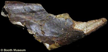
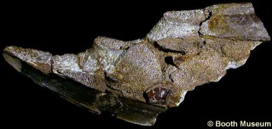
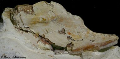
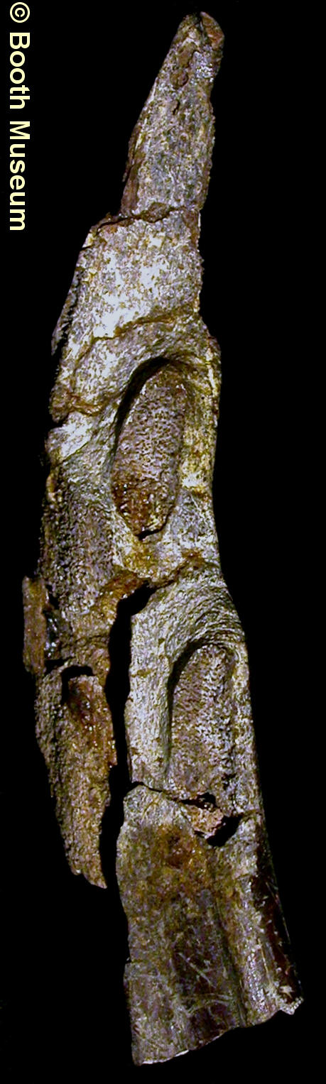
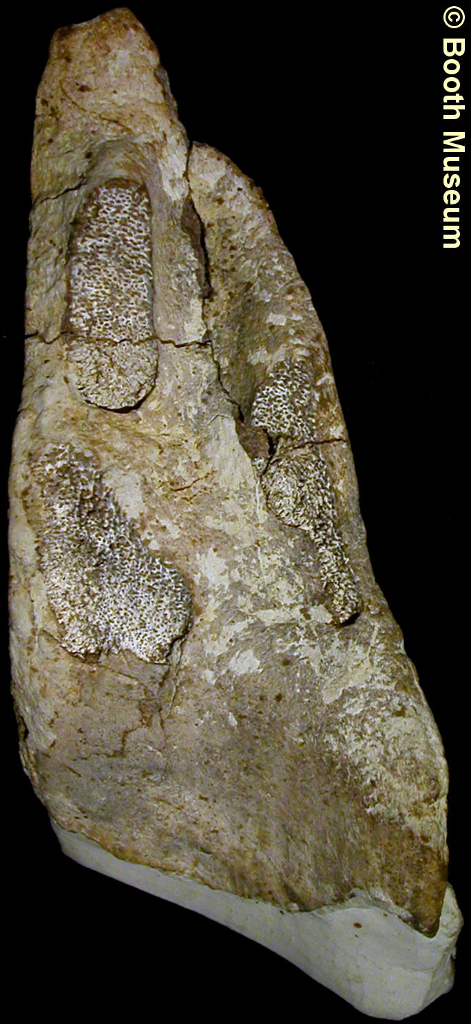
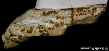
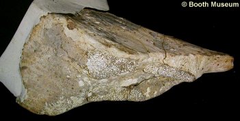
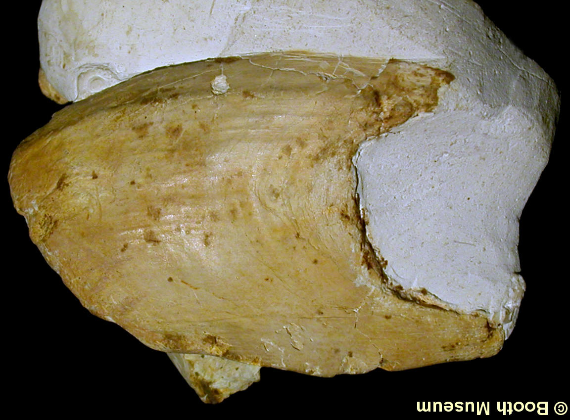
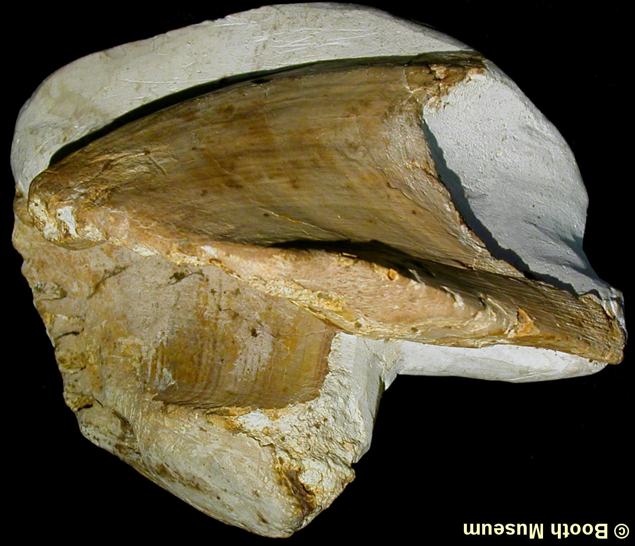
1). Edaphodon mantelli? - right mandibular (from lower jaws); (A) Outer surface, note broken outer surface of beak exposing the inner structure of the dental plate; (B) inner surface. E. mantelli, is characterised as similar to E. sedgwicki (mandibulars with broad symphesial facets and pronounced beak) only smaller and more slender. x1.1, Grey Chalk, West Melbury Marly Chalk, Clayton, near Brighton, West Sussex, Willett Collection, Booth Museum, BMB 007294, by kind permission of John Cooper.
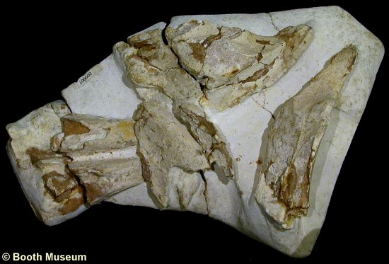
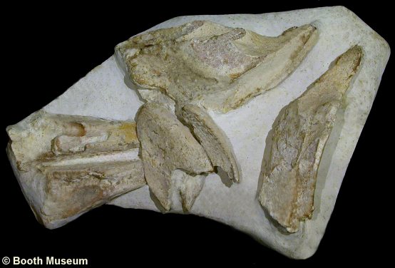
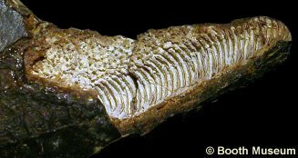
2). Edaphodon sedgwicki: a unique specimen, comprising a full complement of 6 dental plates, from a large individual. Sadly the specimen is much damaged (see Figure 6 for a graphically repaired version). E. sedgwicki is characterised by large dental plates, and mandibulars with pronounced beaks at the front and broad symphesial facets (the articulating surface between mandibulars). x0.65, Lewes, East Sussex, Booth Museum, BMB 009007, by kind permission of John Cooper.
 A A |
 B B |
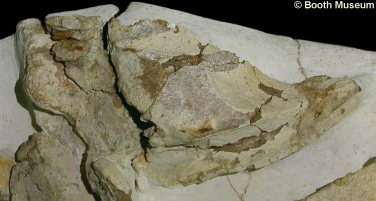
3). Edaphodon sedgwicki - mandibular plates (from the lower jaws) of the above specimen; (A) Inner surface of left mandibular; (B) outer surface of right mandibular. x0.75, Lewes, East Sussex, Booth Museum, BMB 009007, by kind permission of John Cooper.
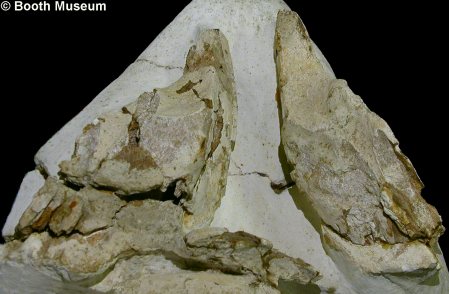
4). Edaphodon sedgwicki - mandibular plates of the above specimen, sitting together as a pair with inner surfaces exposed (equivalent to an internal view of the lower jaws with the front of the head at the top of the image). x0.7, Lewes, East Sussex, Booth Museum, BMB 009007, by kind permission of John Cooper.
 A A |
 B B |
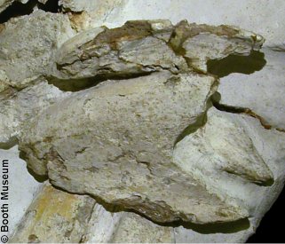
5). Edaphodon sedgwicki - details of the above specimen; (A) Palatine plates (from the rear of of the upper jaws) of the above specimen, sitting together as a pair with inner surfaces exposed (equivalent to an internal view of the rear of the upper jaws, with the front of the head towards the top of the image and the right side of the animal to the left of the image) (x0.95); (B) Vomerine plates (from the front of the upper jaws), resting together as pair, with the outer surface of the left plate foremost (equivalent to an external view of the front of the upper jaws, with the front of the animal to the left of the image) (x1.2). Lewes, East Sussex, Booth Museum, BMB 009007, by kind permission of John Cooper.
6). Edaphodon sedgwicki - graphically repaired overview of the above specimen (compare to Figure 2). x0.65, Lewes, East Sussex, Booth Museum, BMB 009007, by kind permission of John Cooper.
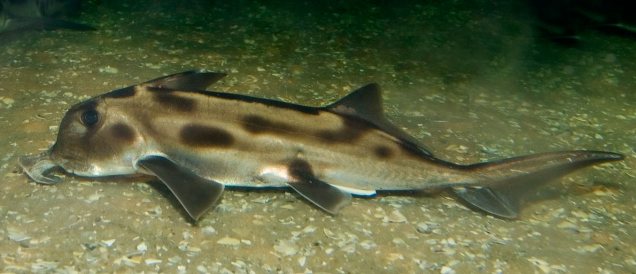
7). Callorhinchus milii (Elephant Shark); A modern Callorhynchid for illustration (image sourced from Wikipedia).
 A A |
 B B |
 C C |
 D D |
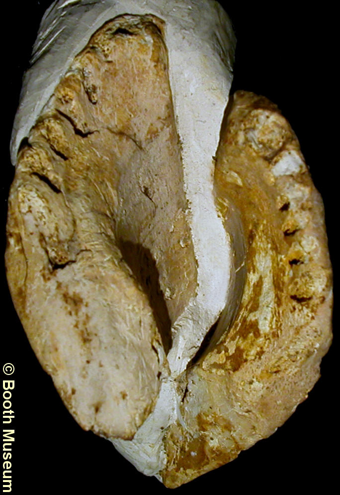
8). View of biting surfaces of mandibulars and palatines; (A) E. mantelli? right mandibular (x1.1) (see Figure 1 for details); (B) E. agassizi? right mandibular (x1.2) (see Figure 9 for details); (C) E. mantelli? left palatine (x1.1) (see Figure 10 for details); (D) E. mantelli? left palatine (x0.9) (see Figure 11 for details). Images courtesy of the Booth Museum, by kind permission of John Cooper.
 A A |
 B B |
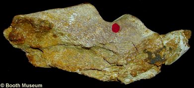
9). Edaphodon agassizi? - right mandibular; Outer surface; (B) Inner surface, note broad symphesial facet and multiple elongate tritors. E. agassiz is characterised by relatively short and stout mandibulars, with relatively under pronounced beaks and relatively narrow symphesial facets. x1.25, Grey Chalk, West Melbury Marly Chalk, Glynde, near Lewes, East Sussex, Willett Collection, Booth Museum, BMB 007290, by kind permission of John Cooper.
 A A |
 B B |
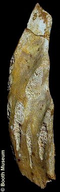
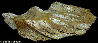
10). Edaphodon mantelli? - left palatine (see also Figure 8C for biting surface); (A) Outer surface (front of head would lie to left of image); (B) Inner surface (front of head woul lie to right of image). x1.2, Grey Chalk, West Melbury Marly Chalk, Clayton, near Brighton, West Sussex, Willett Collection, Booth Museum, BMB 007291, by kind permission of John Cooper.
 A A |
 B B |
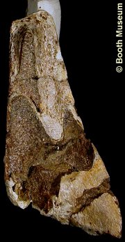
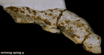
11). Edaphodon mantelli - left palatine right mandibular (see also Figure 8D for biting surface); (A) Outer surface (front of head would lie to left of image); (B) Inner surface (front of head woul lie to right of image). x1.0, Grey Chalk, West Melbury Marly Chalk, Glynde, near Lewes, East Sussex, Willett Collection, Booth Museum, BMB 007293, by kind permission of John Cooper.
 A A |
 B B |
 C C |
12). Edaphodon sp. - pair of vomerine plates; (A) Biting surfaces (equivalent inner view of front of upper jaws, with front of animal towards the top of the image), note multiple tritors along biting edge; (B) Outer surface of left vomerine, front of head to left of image; (C) Oblique view showing outer surface of left vomerine and inner surface of right vomerine, front of head to left of image. x1.0, Grey Chalk, Southerham, near Lewes, East Sussex, Willett Collection, Booth Museum, BMB 007292, by kind permission of John Cooper.
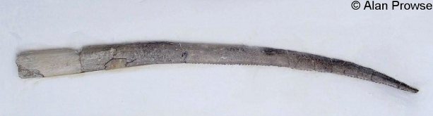
13). Edaphodon sp. - Large fin spine, free of original matrix (plaster base). x0.7, Grey Chalk, Upper Zig Zag Chalk (C. guerangeri zone), between Cow Gap and Holywell, east of Beachy Head, East Sussex, Alan Prowse Collection.
 A A |
 B B |
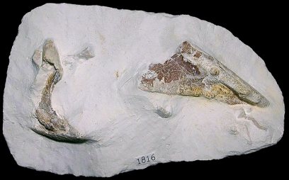
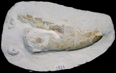
14). Edaphodon mantelli? - associated set of highly degraded dental plates; (A) Palatines?; (B) Right mandibular, outer surface, head to right of image. x1.0, White Chalk, Lewes Nodular Chalk, Seaford Head, East Sussex, Randell collection, RR 1816.
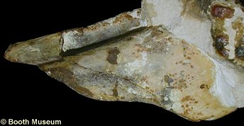
15). Edaphodon mantelli - detail of the specimen in Figure 1. Beak of right mandibular, with outer surface broken away exposing the inner structure of the dental plate. x2.3, Grey Chalk, West Melbury Marly Chalk, Clayton, near Brighton, West Sussex, Willet Collection, Booth Museum, BMB 007294, by kind permission of John Cooper.