|
RHINOCHIMAERIDS [Long-nose Chimaeras] |
Rhinochimaerids are highly scarce in the Chalk and are represented only by the small species Elasmodectes willietti. Mandibular dental plates of E. willetti are highly flattened and shell-like with, rows of minute tritors clustered around the oral margin. A single remarkable specimen preserving much of the originally cartilaginous skeleton is in the collections of the Sedgwick Museum (see below). Dorsal fin spines are straight, with or without barbs or longitudinal ridges.
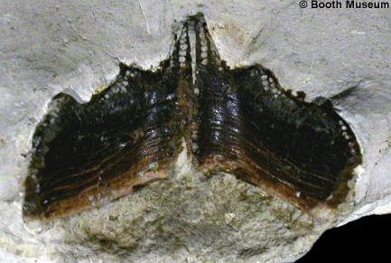
1). Elasmodectes willetti; internal view of the type specimen - a pair of articulated mandibular dental plates (from the lower jaws) (x2.8, Grey Chalk, Southerham, near Lewes, East Sussex, Willett Collection, Booth Museum, BMB 007295, by kind permission of John Cooper).
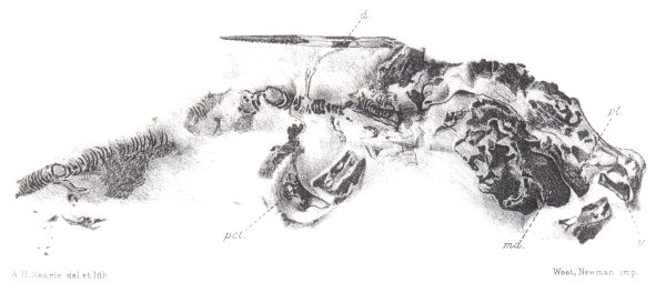
2). Elasmodectes willetti; unique specimen, preserving the cartilaginous skeleton in life articulation (thought to be a female). Note the dorsal fin spine, and the right-hand set of dental plates (md: mandibular plate (from the lower jaw); pl: palatine plate (from the back of the upper jaw); v - vomerine plate (from the front of the upper jaw)). x1.0, Grey Chalk, near Lewes, East Sussex, in the collections of the Sedgwick Museum, Cambridge. Modified from plate XXXIX, figure 4 of Woodward 1902-1912.
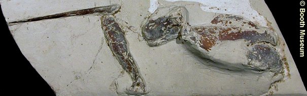
3). Elasmodectes sp. - Dorsal fin spine with portion of pectoral arch and other skeletal elements. x1.3, Grey Chalk, West Melbury Marly Chalk, Clayton, near Brighton, West Sussex, Willett Collection, Booth Museum, BMB 007269, by kind permission of John Cooper.
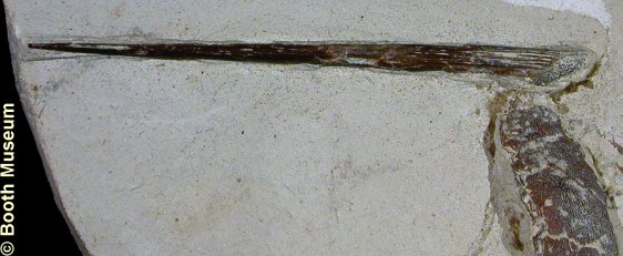
4). Elasmodectes sp. - Detail of above specimen. Dorsal fin spine with portion of pectoral arch. x3.0, Grey Chalk, West Melbury Marly Chalk, Clayton, near Brighton, West Sussex, Willett Collection, Booth Museum, BMB 007269, by kind permission of John Cooper.
|
|
|
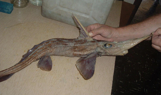
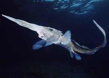
5). (A, B) Images of modern long-nose chimaeras for illustration. Note the prominent fin spine (compare to the specimen in Figure 2) and the jaws protruding beneath the 'nose'. Images courtesy of ImageBaker.com, 2009.