 A A |
 B B |
 C C |
|
HETERODONTIFORMES [Bullhead Sharks]
|
|
|
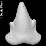
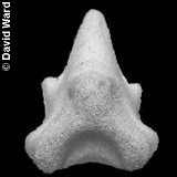
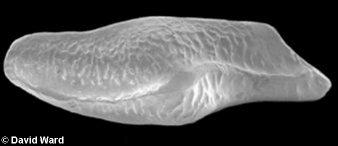
The heterodonts ('differing-teeth') are notable for possessing bony dorsal fin spines, an unusual feature in neoselachians, and markedly different anterior and posterior teeth. The clutching anterior teeth are rounded, labio-lingually flattened cones, with weakly developed, rounded accessory cusps. The crown forms a skirt-like drape over the labial face of the root. The grinding posterior teeth are low and wide, with the cusps reduced and merged into a long, low, rounded keel, ornamented with weak ridges perpendicular to the cutting edge. There are a number of exceptionally preserved Chalk specimens in the major museum collections, preserving the articulated cartilaginous skeleton, some of which are figured below. Isolated teeth are uncommon in the field.
|
A |
B |
C |
1). Heterodontus - Representative isolated Heterodontiform teeth from the Albian Gault Clay of Folkestone, David Ward Collection: (A, B) Heterodontus canaliculatus - (A) Labial and (B) lingual views of an anterior tooth (x19); (C) Heterodontus sp. - crown/oral view of a latero-posterior tooth (x21). Images by kind permission of David Ward. See also www.gaultammonite.co.uk [Fossils of the Gault Clay].
|
|
 B B |
 C C |
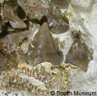
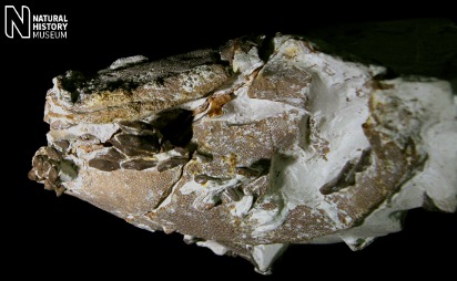
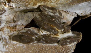
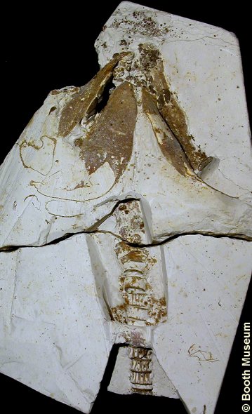
2). Heterodontus canaliculatus - Ventral (underside) view of a remarkable specimen, with robust and three dimensional calcareous preservation of the originally cartilaginous skeletal elements of the head and anterior portion of the trunk. (A) (x1.0, White Chalk, Turonian, Malling, near Lewes, East Sussex, Willett Collection, Booth Museum, BMB 007329, by kind permission of John Cooper); (B) Anterior teeth of the upper jaws (palatoquadrate) (x11, detail of Figure 1A); (C) Lateroposterior teeth of the upper jaws (x12, detail of Figure 6A).
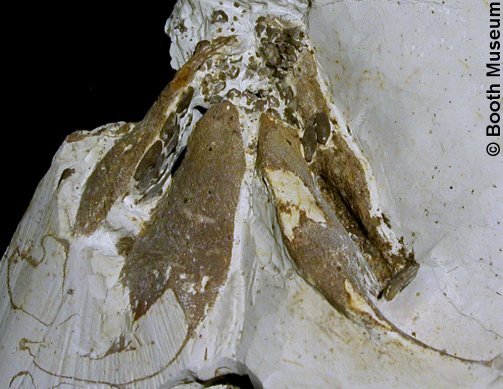
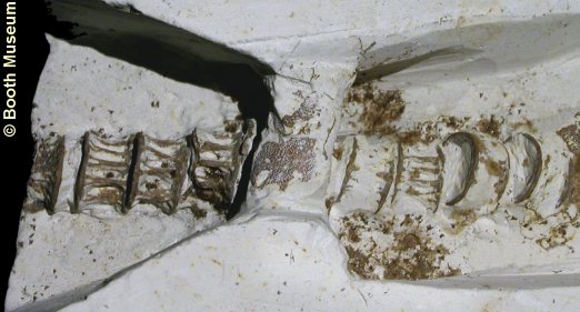
3). Heterodontus canaliculatus - Detail of the above specimen. Ventral (underside) view of the head, showing lower and upper jaws (mandibular arch) with teeth in situ. The change in the form of the teeth from the back of the jaws (broad and blunt) (latero-posterior teeth) to the front of the jaws (narrow and sharp) (anterior teeth) is clearly apparent (x1.5).
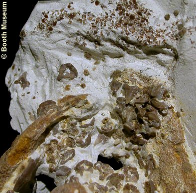
4). Heterodontus canaliculatus - Detail of the above specimen, front of the animal towards the top of the image. Ventral (underside) view of the front of the jaws, showing the abrupt change from broad and blunt lateral teeth (bottom left of the image) to narrow and sharp anterior teeth. The material at the top of the image is a concentration of placoid scales from the shagreen (skin) at the snout of the animal (x3.3).
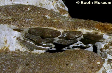
5). Heterodontus canaliculatus - Detail of the above specimen, front of the animal towards the right of the image. Ventral (underside) view of the right hand set of jaws. The biting surface of broad and blunt latero-posterior teeth of the upper right hand jaw (palatoquarate) is apparent (x2.7).
6). Heterodontus canaliculatus - Detail of the above specimen, front of the animal towards the right of the image. Ventral (underside) view of the vertebral column and the overlying pectoral girdle (chest bone; only partially exposed) (x2.6).
|
|
 B B |
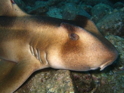
7). Heterodontus - Modern Heterodontids for illustration: (A) Horn Shark (Heterodontus francisci) - note prominent dorsal fins supported by bony fin spines; (B) Crested Bullhead Shark (Heterodontus galeatus). (images sourced from Wikipedia).
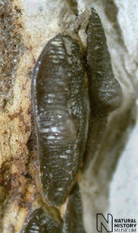
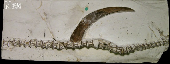
8). Heterodontus canaliculatus - Series of vertebrae with both dorsal fin spines (x0.8, Grey Chalk, Glynde, Sussex, NHMUK (British Museum (Natural History) London) PV P49734). Image © 2012 The Natural History Museum, by kind permission.
9). Heterodontus canaliculatus - Series of vertebrae with fin spines (front of individual to the right) (x0.9, Booth Museum, BMB 008555), by kind permission of John Cooper).
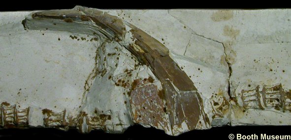
10). Heterodontus canaliculatus - Series of verterae with fin spine (x1.9, Booth Museum, BMB 008555), by kind permission of John Cooper).
|
|
 B B |
11). Heterodontus canaliculatus - (A) Ventral (x1) and (B) left lateral (x1.2) views of a large set of three-dimensional jaws with associated teeth (Grey Chalk, Lewes, Sussex, NHMUK (British Museum (Natural History) London) PV P20123). Images © 2012 The Natural History Museum, by kind permission.
|
|
 B B |
12). Heterodontus canaliculatus - Details of the above specimen: (A) Anterior tip of the lower jaws (Meckel's cartilage) with worn and blunted anterior teeth in crown/oral view (x4); (B) Crown/oral views of semi-articulated latero-posterior teeth from opposing jaws (x3.5) (Grey Chalk, Lewes, Sussex, NHMUK (British Museum (Natural History) London) PV P20123). Images © 2012 The Natural History Museum, by kind permission.
|
|
|
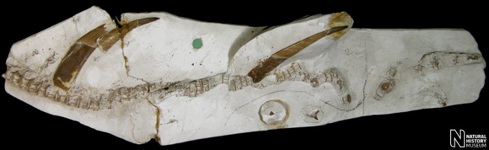
13). Heterodontus canaliculatus - Part and counterpart of a very well preserved and articulated set of jaws (mandibular arch) with teeth: (A) ventral (as if from the underside) view of the right hand side of the head, and anterior most portion of the left hand side of the head; latero-posterior teeth of the right hand upper jaw have root surface exposed; (B) dorsal (as if from the upper side) view of the right hand side of the head; latero-posterior teeth (biting surface exposed) and anterior teeth are apparently from the upper jaw (palatoquadrate) (x1.6, Grey Chalk(?), Guilford, Surrey, NHMUK (British Museum (Natural History) London) PV P49735). Images © 2012 The Natural History Museum, by kind permission.
|
|
|
14). Heterodontus canaliculatus - Details of the above specimen: Anterior tip of the upper jaws (palatoquadrate) with anterior teeth (x8); (B) Crown/oral views of semi-articulated latero-posterior teeth of the left upper jaw (x6.5) (Grey Chalk(?), Guilford, Surrey, NHMUK (British Museum (Natural History) London) PV P49735). Images © 2012 The Natural History Museum, by kind permission.
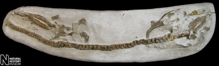
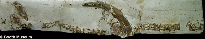
15). Heterodontus sp.? - An unusual specimen, comprising a large series of articulated vertabrae with asociated skeletal elements (x0.55, Grey Chalk, Dover, Kent, NHMUK (British Museum (Natural History) London) PV P49079). Image © 2012 The Natural History Museum, by kind permission.
|
|
|
16). Heterodontus sp. - (A,B) Part and counterpart of the largely articulated anterior portion of the body (x1.1, Grey Chalk, Kent, NHMUK (British Museum (Natural History) London) PV P38550). Images © 2012 The Natural History Museum, by kind permission.
|
|
|
17). Heterodontus canaliculatus - (A) Ventral and (B) right lateral view of the a set of articulated three dimensional jaws (x1.75, Grey Chalk, Lewes, Kent, NHMUK (British Museum (Natural History) London) PV P4471). Images © 2012 The Natural History Museum, by kind permission.
|
|
|
18). Heterodontus canaliculatus - A semi-unique example of jaw preservation from the 'Upper' White Chalk: (A) Ventral view of the lower jaws; (B) Dorsal view of the specimen, showing the vertebrae joining the back of the head (x1.1, White Chalk (post-Turonian), NHMUK (British Museum (Natural History) London) PV P39061). Images © 2012 The Natural History Museum, by kind permission.
|
|
|
20). Heterodontus canaliculatus - Dorsal spines with associated skeletal elements: (A) x1.5, Chalk, Lewes, Sussex, NHMUK (British Museum (Natural History) London) PV P4084; (B) x1.5, Chalk, Sussex, NHMUK (British Museum (Natural History) London) PV P1294. Images © 2012 The Natural History Museum, by kind permission.
|
|
|
21). Heterodontus sp. - Isolated dorsal spines: (A) x2.6, White Chalk, Norwich, Norfolk, NHMUK (British Museum (Natural History) London) PV P48949; (B) x1.4, Chalk, Lewes, Sussex, NHMUK (British Museum (Natural History) London) PV P4080. Images © 2012 The Natural History Museum, by kind permission.
 A
A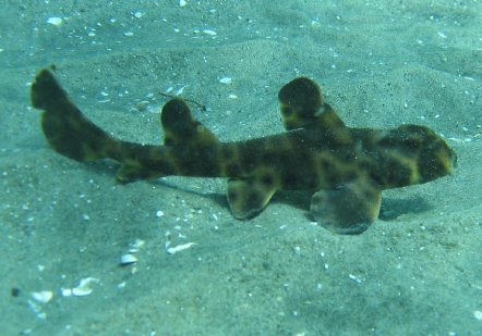 A
A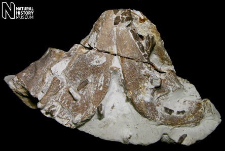 A
A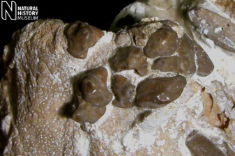 A
A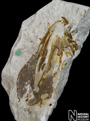 A
A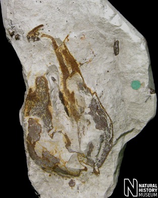 B
B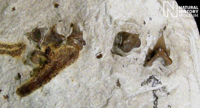 A
A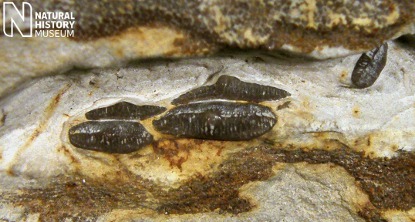 B
B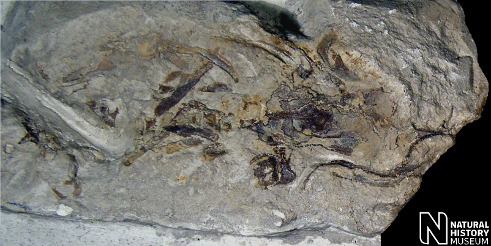 A
A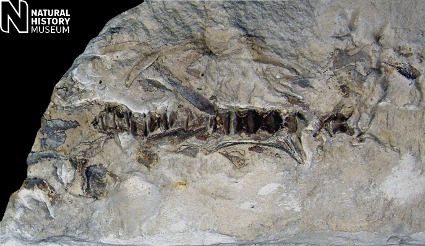 B
B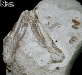 A
A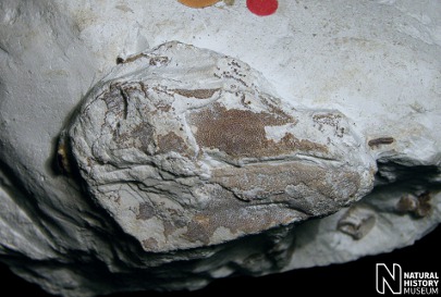 B
B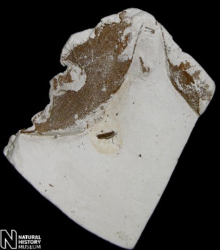 A
A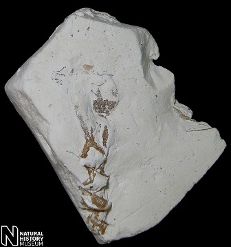 B
B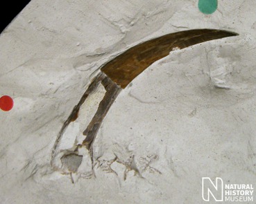 A
A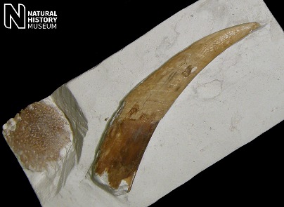 B
B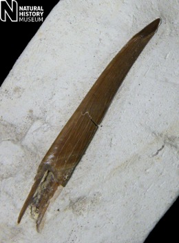 A
A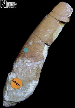 B
B