|
Squatiniformes [Angel Sharks] |
|
|
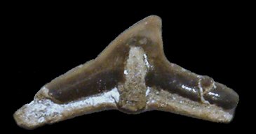
Angel Sharks possess a flattened ray-like body plan, and a large number of small but distinctive clutching / grasping teeth. These are low and wide, with a simple, central triangular main cusp and a low root with a broad, flattened base. There are no accessory cusps, instead there are long, low areas of crown either side of the main cusp. A pendant-like extension of the crown hangs down over the labial face of the root directly below the main cusp. Anterior-most teeth are of conservative form, comprising a simple, narrow and distally-curved main cusp, taller than wide. A single exceptionally preserved individual is recorded (figured below). Isolated teeth are not abundant in the field.
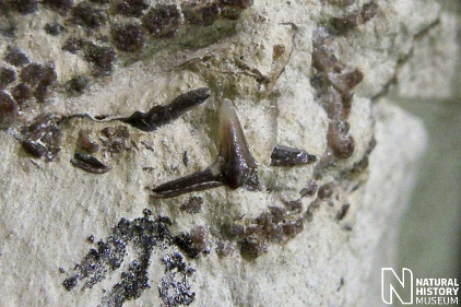
1). Squatina sp. - (A) Labial view of an isolated latero-posterior tooth, with a prominent medial 'rod' of enamaloid bridging the root and the base of the main cusp (x12, Seaford Chalk, St Margarets, Kent, Randell Collection RR1389).
 A
A |
 B
B |
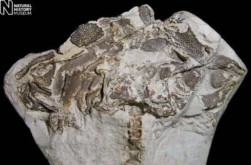
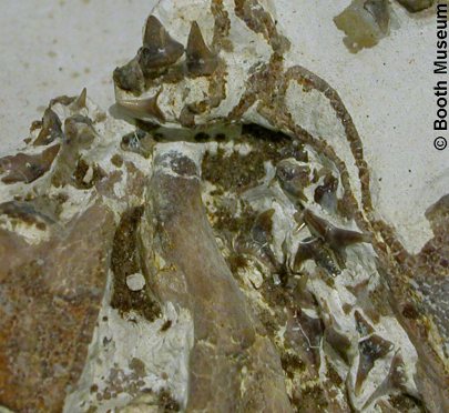
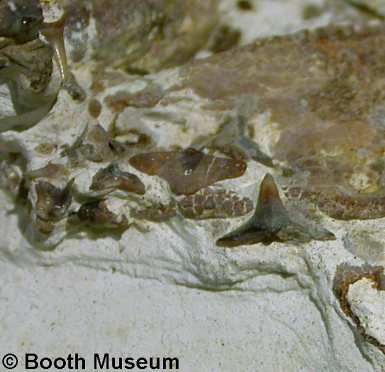
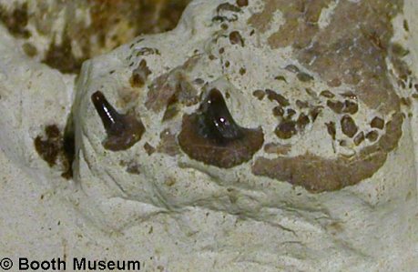
2). Squatina cranei; details of the type specimen displaying in situ teeth (Grey Chalk, Clayton, near Brighton, Sussex, Willett Collection, Booth Museum, BMB 007330, by kind permission of John Cooper); (A) Ventral-view detail of the anterior portion of the jaws, displaying anterior teeth of the palatoquadrate (upper jaws) (top centre) and latero-posterior teeth of the upper jaws (centre right and bottom right). The dorsal (under-) surface of the Meckel's cartilage (lower jaws) is seen (bottom left quarter of mage) framed by the teeth of the upper jaws, and the upper jaws themselves (right of image) (x4.5); (B) Lateral view of the anteriormost portion of the jaws and anterior teeth, underside-up (x7).
 A
A |
 B
B |
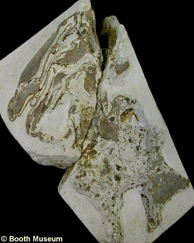
3). Squatina cranei - (A) Part and (B) counterpart of the exceptionally preserved type specimen figured above, which displays robust calcification of the originally cartilaginous skeletal elements of the head. The front of the animal is towards the top of the image in both views; (A) Dorsal view of the head and anterior portion of the trunk, including a possible remnant of the right pectoral fin (bottom right of the image)(x1.2); (B) Ventral (underside) view of the head (mandibular and hyoid arches), displaying the undersurface of the lower jaws (Meckel's cartilage) in life position, framed within the upper jaws (palatpquadrate), both retaining their complement of teeth (x1.7).
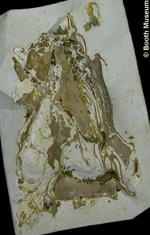
4). Squatina cranei - Overview of remarkably preserved specimen, with an articulated vertebral column (evidently complete at least as far as the pectoral arch as preserved) and near complete complement of cartilaginous elements of the head. A small number of associated teeth are also seen (x1.2, Grey Chalk, Halling, Kent, NHMUK (British Museum (Natural History) London) PV P12213). Image © 2012 The Natural History Museum, by kind permission.
|
|
|
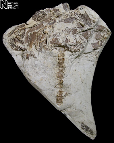
5). Squatina cranei - Details of (A) the cartilaginous elements of the head and (B) labial
view of an anterior tooth ((A) x2.3, (B) x15, Grey
Chalk, Halling, Kent, NHMUK (British
Museum (Natural History) London)
PV P12213). Images © 2012 The Natural History Museum, by kind
permission.
 A
A |
 B B |
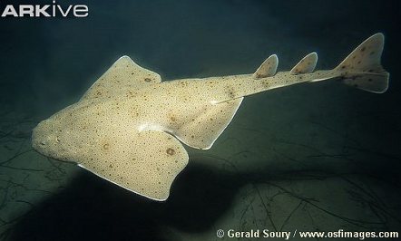
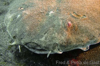
6). Modern angel sharks for illustration; (A) dorsal view of a Pacific Angel Shark (Squatina californica) (image sourced from ARKive, © Gerald Soury / www.osfimages.com); (B) Head of an Angel Shark (Squatina squatina) (image sourced from Onderwaterfoto's van Fred en Petra de Gast).
 A
A |
 B
B |
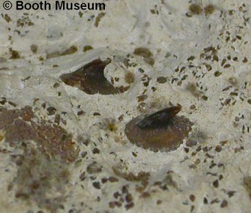
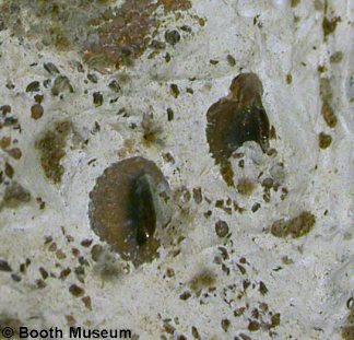
7). Squatina cranei; details of the type specimen figured above (Figure 2); (A, B) dermal denticles and other elements embedded in the skin of the animal in life (A, x6.5)(B, x6.5).
8). Squatina cranei; detail of the type specimen figured above (Figure 2) showing dermal denticles resting against a calcified formerly cartilaginous skeletal element from the back of the head (x9).
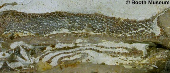
9). Squatina cranei; details of the type specimen figured above (Figure 2) showing the calcified formerly cartilaginous left upper jaw broken open to reveal the 'cellular' structure (x5).