HOMEPAGE

FOSSILS VERTEBRATES CHONDRICHTHYES TERMINOLOGY REFERENCES LINKS


SYNECHODONTIFORMES
Synechodus
(Woodward, 1888)
Paraorthacodus (Glückman, 1957)


Though teeth of these relatively 'primitive' neoselachians are uncommon in the field, the synechodonts are well represented amongst the small number of exceptionally preserved shark specimens from the Chalk.  Examples of articulated specimens exhibiting preservation of the cartilage are figure below.

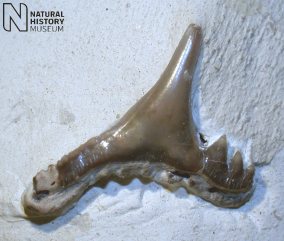
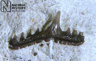
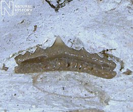
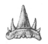
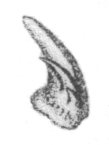
The teeth are distinctive, and several species are recognised.  Synechodus teeth, known from the Grey Chalk, are wide and low, with a very shallow root bearing a series of prominent basal notches.  The crown is low, but with a prominent central main cusp in anterior teeth, and a series of thorn-like accessory cusps.  Paraorthacodus teeth, known from the Campanian, are less broad and have a crown divided into a large, tall, distally curved central main cusp, directly sided by a small number of tall, narrow accessory cusps.


 A  B

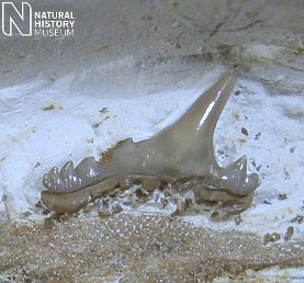
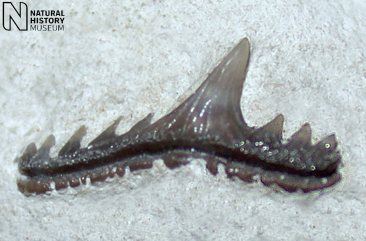
1).  Individual teeth; (A) Synechodus dubrisiensis (x9.0, Grey Chalk, Dover, Kent, NMHUK (British Museum (Natural History) London) PV P47287); (B) Synechodus nitidus, isolated tooth - note the arched nature of the root, and the smooth swollen labial and lingual surfaces to the base of the main cusp (x6.0, Grey Chalk, Wouldham, near Rochester, Kent, NMHUK PV P10228).  Images courtesy of Charlie Underwood, © 2008 The Natural History Museum, by kind permission.


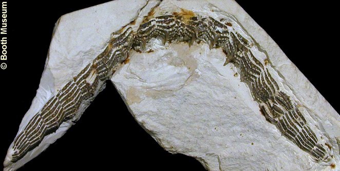
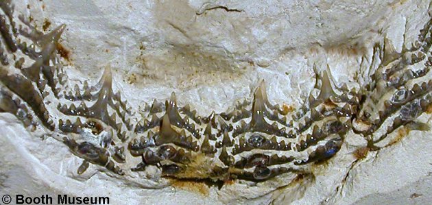
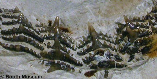
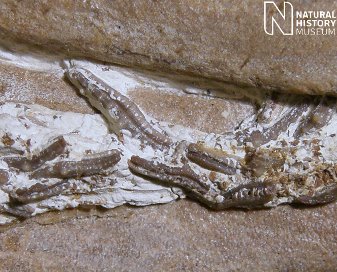
2).  Synechodus dubrisiensis; a unique specimen of a single jaw, lacking the cartilaginous jaw parts, but preserving the complete dentition in perfect articulation (x2.7, Grey Chalk, Sussex, Willett Collection, Booth Museum, BMB 008523, by kind permission of John Cooper).


3).  Synechodus dubrisiensis; detail of the anterior-most portion of the above specimen; note the two central rows of miniscule symphysial teeth (x6, Grey Chalk, Sussex, Willett Collection, Booth Museum, BMB 008523, by kind permission of John Cooper).).


4).  Synechodus dubrisiensis; lateral portion of the above specimen (x7, Grey Chalk, Sussex, Willett Collection, Booth Museum, BMB 008523, by kind permission of John Cooper).).


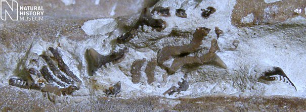
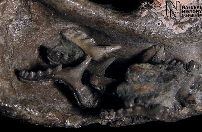
5).  Synechodus dubrisiensis; detail of the above specimen (x8).

  
A    B
 
 C
6).  Synechodus dubrisiensis - exceptional specimen displaying full three-dimensional preservation of cartilaginous skeletal elements of the jaws (mandibular and hyoid arches) with teeth retained in-situ; (A) Underside (ventral) view, (B) detail of anterior teeth, and (C) detail of latero-posterior teeth ((A) x1.6, (B) x4.0, (C) x3.0), Chalk, Kent, NHMUK (British Museum (Natural History) London) PV P41675.  Images © 2011 The Natural History Museum, by kind permission.

--

A  B

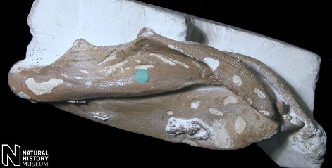
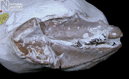
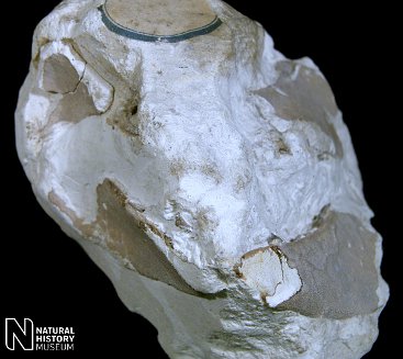
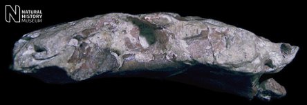
7).  Synechodus dubrisiensis - (A) left and (B) right lateral views of the specimen displaying three-dimensional preservation of cartilaginous skeletal elements of the jaws with teeth retained in-situ; (A, B x2.5, Chalk, Kent, NHMUK (British Museum (Natural History) London) PV P41675.  Images © 2011 The Natural History Museum, by kind permission.

--


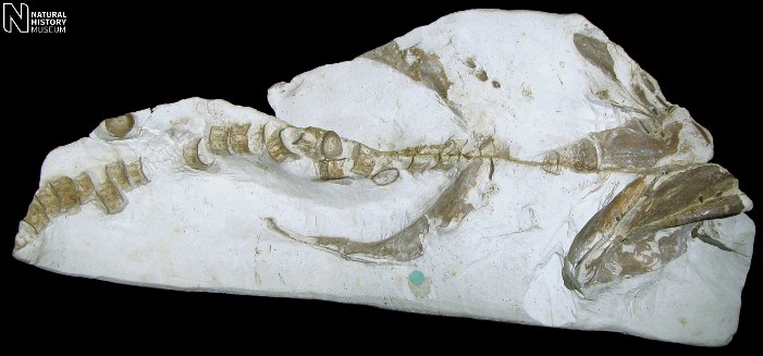
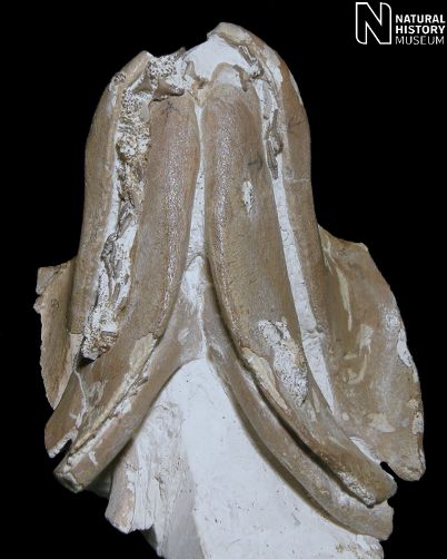
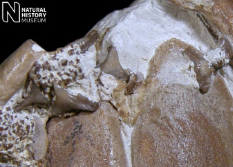
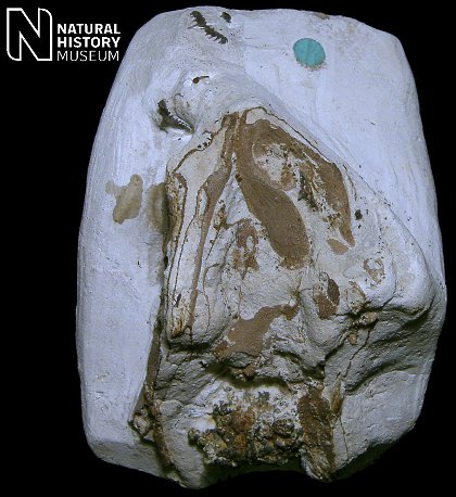
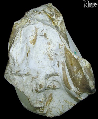
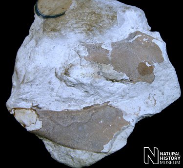
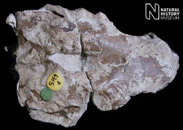
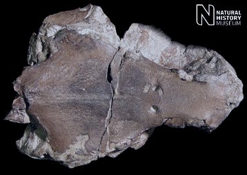
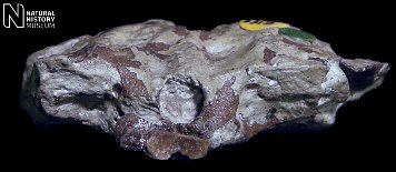
8). Synechodus dubrisiensis - Ventral (underside) view of a partial anterior portion of an exceptionally preserved specimen (front of animal to the right) displaying jaws (mandibular arch), hyoid arch, chondrocranium, vertebrae and pectoral arch (x1.0, Zig Zag Chalk, Burham, Kent, NHMUK (British Museum (Natural History) London) PV P49032.  Image © 2011 The Natural History Museum, by kind permission.

--

A B

9).  Synechodus dubrisiensis(A) Lateral (right-side) and (B) ventral (underside) views of the holotype - small set of jaws (mandibular arch) with teeth (A,B x1.5, Zig Zag Chalk, Dover, NHMUK (British Museum (Natural History) London) PV P36908.  Images © 2011 The Natural History Museum, by kind permission.

--

10).  Synechodus dubrisiensisDetail of the holotype retaining latero-posterior teeth in-situ (x4.5, Zig Zag Chalk, Dover, NHMUK (British Museum (Natural History) London) PV P36908).  Images © 2011 The Natural History Museum, by kind permission.

--

A    B
 
 C

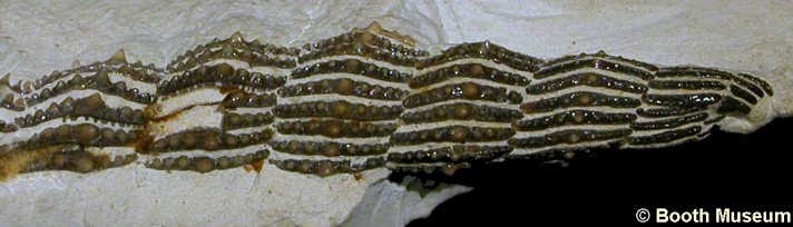
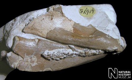
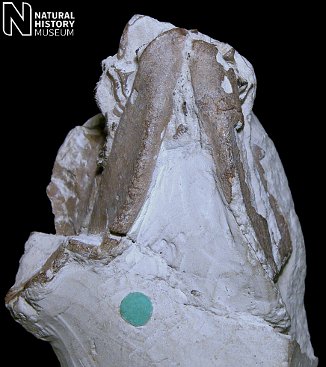
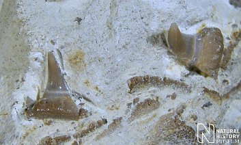
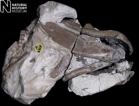
11).  Synechodus dubrisiensis: (A) Ventral (underside) view of an exceptional specimen preserving the cartilaginous skeletal elements of the head; the front of the animal is to the top, most of the exposed material is the Meckel's cartilage (x2.0); (B) Detail of a lateral tooth (x10.0); (C) Detail of a posterior tooth (x10.0).  (Grey Chalk, Dover, Kent, NHMUK (British Museum (Natural History) London) PV P47287).  Images © 2011 The Natural History Museum, by kind permission.

--

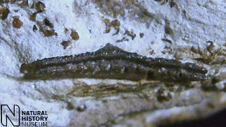
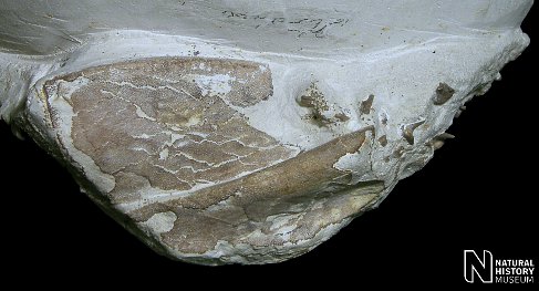
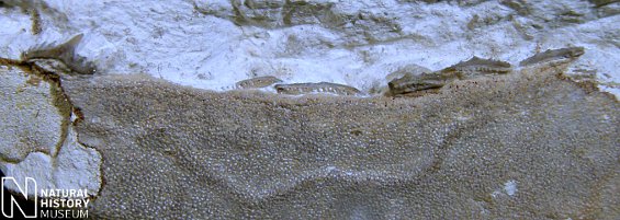
12).  Synechodus dubrisiensis: Detail of an exceptionally preserved specimen retaining dermal denticles (x15.0, Grey Chalk, Dover, Kent, NHMUK (British Museum (Natural History) London) PV P47287).  Image © 2011 The Natural History Museum, by kind permission.

--

A    B
 
 C
13).  Synechodus nitidus - exceptionally preserved holotype specimen displaying three-dimensional preservation of cartilaginous skeletal elements of the jaws (mandibular and hyoid arches) with teeth retained: (A) Underside (ventral) view (x1.1), (B) Detail of anterior teeth (x6.0); (C) Detail of latero-posterior tooth (x8.0).  (Grey Chalk, Snodland, Kent, NHMUK (British Museum (Natural History) London) PV P1295).  Images © 2011 The Natural History Museum, by kind permission.

--

A

 B

14).  Synechodus nitidus - further views of the holotype specimen displaying: (A) Rightt-lateral view of the jaws (x1.0); (B) detail of an anterio-lateral tooth (x6.0).  (Grey Chalk, Snodland, Kent, NHMUK (British Museum (Natural History) London) PV P1295).  Images © 2011 The Natural History Museum, by kind permission.

--

A  B
15).  Synechodus dubrisiensis - damaged remains of a very well preserved specimen with 3-dimensional jaws and in-situ teeth: (A) left lateral and (B) anterior (dorsal to top of image) views of the jaws (x,1.3, Grey Chalk, Cambridge, NHMUK (British Museum (Natural History) London) PV P7129).  Images © 2011 The Natural History Museum, by kind permission.

--

16).  Synechodus dubrisiensis - detail of well preserved left lower jaw (Meckel's cartilage) with associated latmeral and posterior teeth in life arrangement (anterior to the left) (x,4.0, Grey Chalk, Cambridge, NHMUK (British Museum (Natural History) London) PV P7129).  Images © 2011 The Natural History Museum, by kind permission.

--

A  B
17).  Synechodus dubrisiensis - Exceptionally specimen preserving a full compliment of cartilaginous skeletal elements of the head and with associated teeth (see below for chondrocranium): (A) Underside (ventral) view, anterior to the right (x1.6); (B) Detail of the inner surface of the anterior region of the right upper jaw (palatoquadrate) with anterior teeth in life arangement (x8.0).  Grey Chalk, FolkestoneNHMUK (British Museum (Natural History) London) PV P6315).  Images © 2011 The Natural History Museum, by kind permission.

-

A  B
18).  Synechodus dubrisiensis - Semi-unique example of a shark chondrocranium from the British Chalk: (A) Dorsal and (B) ventral views, anterior to the right (x1.8).  Grey Chalk, FolkestoneNHMUK (British Museum (Natural History) London) PV P6315).  Images © 2011 The Natural History Museum, by kind permission.

--

A  B
19).  Synechodus dubrisiensis - Semi-unique example of a shark chondrocranium from the British Chalk: (A) right lateral (anterior to right) Dorsal and (B) posterior (dorsal to top) views, anterior to the right (x2.0).  Grey Chalk, FolkestoneNHMUK (British Museum (Natural History) London) PV P6315).  Images © 2011 The Natural History Museum, by kind permission.

--

 A  B

20).  Paraorthacodus recurvus; (A) Lingual and (B) side views of an isolated tooth (x3.0, Zig Zag Chalk, Norwich, NMHUK (British Museum (Natural History) London) PV P48953).  Images (A, B) taken from Woodward (1902-1912, Plate XLVI, figures 8 and 8a respectively.