|
NEOSELACHII INCERTAE SEDIS
Ptychodus (Agassiz, 1835) |
|
|
The distinctive
crushing / grinding teeth of Ptychodus are amongst the most familiar of
Chalk fossils, but the taxonomic placement of the Ptychodontid sharks is an
ongoing point of controversy. They
possess a problematic combination of characters which suggest a number of
non-compatible affinities - their tooth microstructure like
Hybodontids,
their tooth morphology like rays (Batomorphii),
their jaw structure like
Heterodontiformes,
and their vertebrae like
Lamniformes. They are widely thought of as rays by amateur
collectors, though the academic community seems to have dismissed this option,
largely viewing them as relatively
'primitive' euselachians, close to the Hybodontids. However, recent
work based on vertebra from well preserved US material has concluded that they
are
neoselachians (Everhart & Caggiano, 2004).
Spectacular complete and
articulated sets of Ptychodus teeth are encountered occasionally in the
US. Associations of teeth and vertebrae are also known from the UK, as
well as exceptionally preserved jaw sets retaining the teeth (see below).
Generally teeth comprise of a large robust crown,
approximating a rounded square in plan view, with a blocky rounded-cube shaped
root tucked underneath. Tooth form
is highly variable and a perhaps surprising number of
Ptychodus species are recognised from the Chalk, each defined on
overall tooth form. The main species concepts can be summarised as
follows:
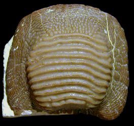
- P. decurrens –
This is the common form from the
Grey Chalk and lowest
White Chalk
and is highly variable.
Flattened to bulbous in
lingual
profile, but without a depressed marginal area. Crown
crossed by a large number of transverse ridges, often finely bifurcated
towards the margins, and passing diffusely into a narrow and poorly defined
marginal area with granular ornament. A var. depressus
form is recognised with a flattened central area, and a var.
multistriatus form where ridges join at the ends to form concentric
rings. [Cenomanian
to
Turonian].
- P. marginalis –
Central area is broad, flattened and moderately elevated in lingual profile,
with a broad depressed marginal area. The central area is crossed by a
large number of longitudinal ridges. [White Chalk: Turonian to
Coniacian].
- P. mammillaris –
Central area is narrow, rounded and highly elevated in lingual profile, with a
depressed marginal area. The central area is crossed by a small number
of longitudinal ridges. [White Chalk: Turonian
to Coniacian].
- P. rugosus –
Like P. mammilaris, but with poorly defined ridges on the central area
passing into a rugose ('lumpy') ornament. [White Chalk: Coniacian to
Santonian].
- P. latissimus – Low rounded profile in
lingual view. The central area is crown crossed by a relatively small number of
highly pronounced longitudinal ridges, triangular in section. Relatively
wide marginal area, not depressed, and with fine granular ornament. P. 'dixoni'
form with finer ridges to central area, P. 'paucisulcatus' form with very
few ridges. [White and grey Chalk: Cenomanian to Santonian].
- P. polygyrus -
Low in profile, with broad, highly flattened central area, and typically very
narrow marginal area. Central area ornamented by a relatively small
number of pronounced longitudinal ridges, often joined to one another at the
ends giving concentric ringed effect. [White Chalk: Coniacian to
Santonian].
- P. concentricus - Like P.
polygyrus, but with a highly domed central area. Scarce in the UK.
[Grey Chalk: Cenomanian].
- P. oweni -
Low and rounded in lingual profile, with no depression of the narrow marginal
area. Ridged ornament of the central area passes into a finer ridged
ornament of the marginal area, all loosely arranged in a part-radial,
part-longitudinal fashion. [Grey Chalk: Cenomanian].
- P. mortoni
- A US form, virtually unknown from the UK. Rounded to sub-triangular in
side profile. Central area bears a radial ornament of coarse ridges
converging towards a central apex, with a broad margin with granular ornament.
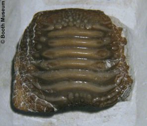
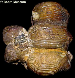
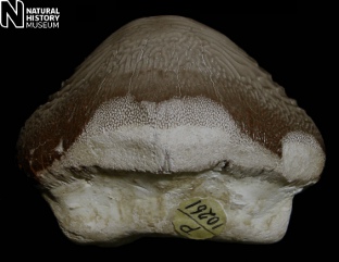
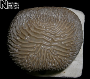
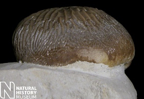
1).
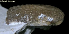
(A,B)
Ptychodus cf. marginalis - (A) oral (biting surface) and (B) side views
of a large tooth - note broad margin and raised central area (x1.4,
Booth Museum, BMB 016994, by kind permission
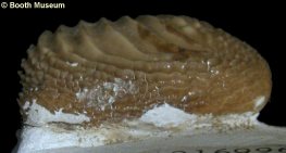
of John Cooper); (C, D) Ptychodus latissimus; (C) oral (biting surface)
and (D) side views of a large tooth - note weakly raised central area
crossed by a relatively small number of broad ridges (x1.4,
Booth Museum, BMB 016998, by kind permission
of John Cooper).
|
 A A
|
 B B |
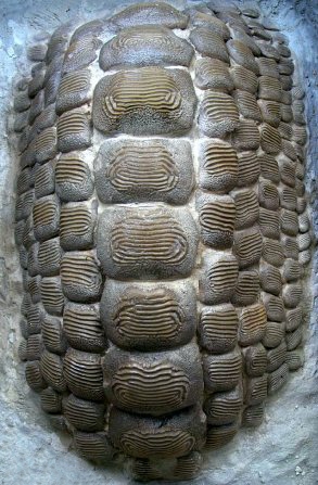
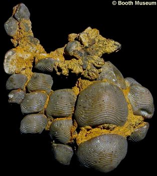
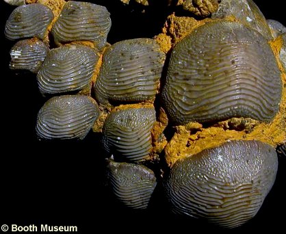
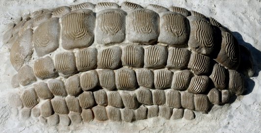
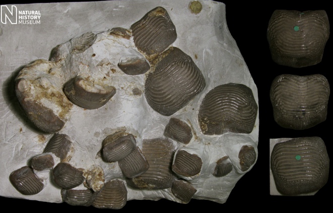
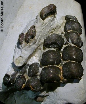
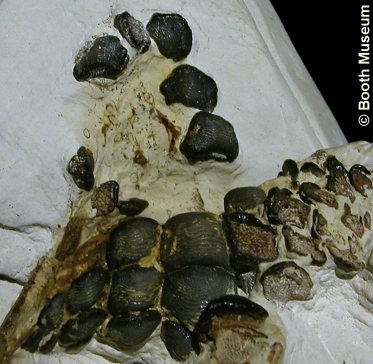
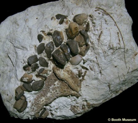
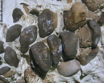
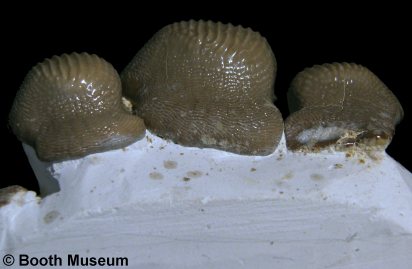
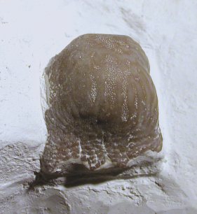
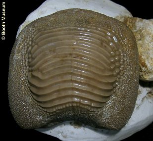
2).
Ptychodus decurrens; oral (biting surface) views of an exceptional
articulated portion of the lower dentition, fused by pyrite / marcasite.
The front of the animal presumably lies towards the top of the image. The
single central row of larger teeth indicates that this is the lower dentition (Grey Chalk of
Southerham, near Lewes, Sussex, Willett Collection,
Booth Museum, BMB 007331, by kind permission
of John Cooper); (A) general view of specimen (x0.9); (B) detail, with central
row of enlarged teeth on the right (x1.5).
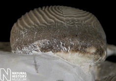
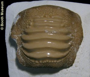
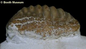
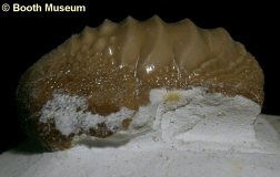
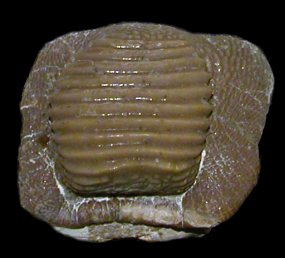
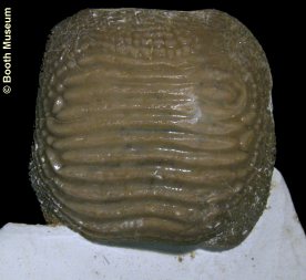
3). (A,B)
Ptychodus decurrens - (A) oral (biting surface) and (B) side views of a
typical tooth - note narrow, indistinct margin and slightly rounded character (x1.6,
Booth Museum, BMB 016996, by kind permission
of John Cooper); (C, D) Ptychodus cf. polygyrus - (C) oral (biting surface)
and (D) side views of a large tooth
- note flattened profile, and central ridges looped-back and joined at
through the margins giving a concentric pattern (x1.6,
Booth Museum, BMB
011247,
by kind permission of John Cooper).
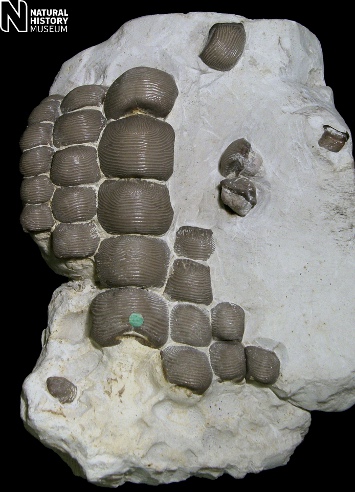
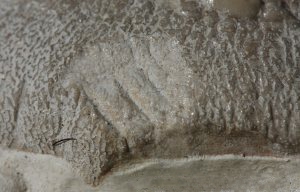
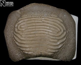
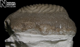
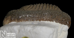
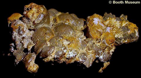
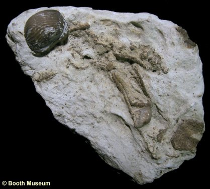
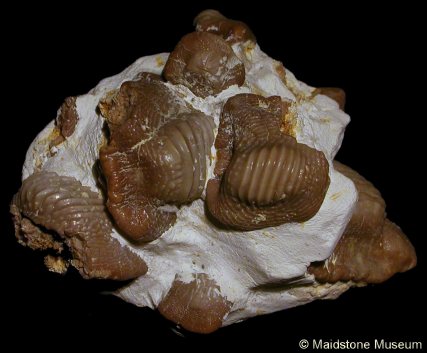
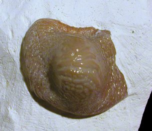
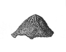
4).
Ptychodus decurrens - partial
articulated palate from the upper jaw. Tooth impressions at the
base of the specimen indicate that significantly more teeth were
originally present (x1.0, Grey
Chalk, Maidstone, Kent, NHMUK (British
Museum (Natural History) London)
PV P40056). Image © 2012 The Natural History Museum, by kind
permission.
 A A |
 B B |
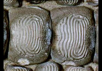
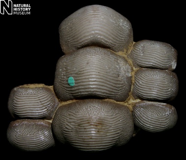
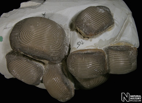
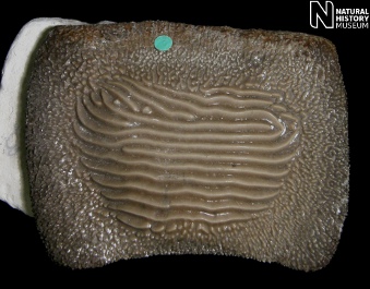
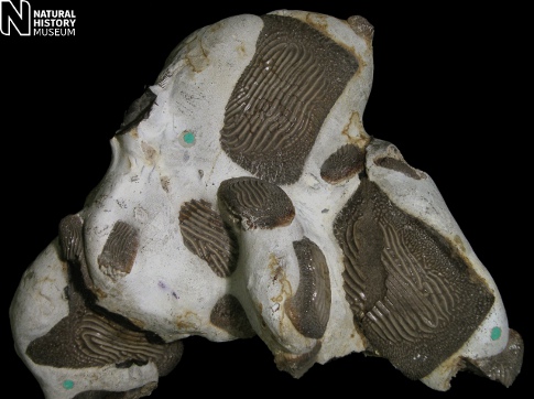
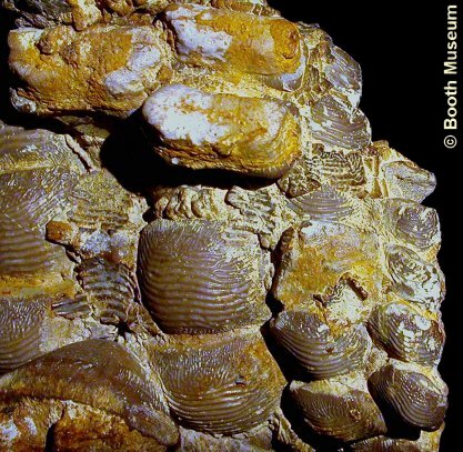
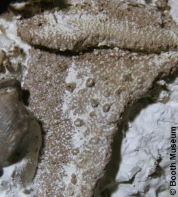
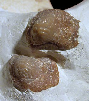
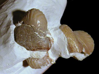
5).
Ptychodus decurrens - articulated palate fragments from the upper jaw. (A) x1.6, Grey
Chalk, Merstham, Surrey, NHMUK (British
Museum (Natural History) London)
PV P12830; (B) x2.7, Grey
Chalk, Halling, Kent, NHMUK (British
Museum (Natural History) London)
PV P38564. Images © 2012 The Natural History Museum, by kind
permission.
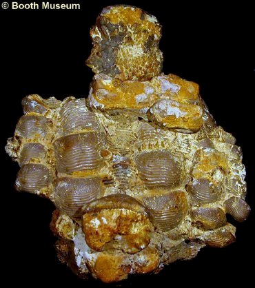
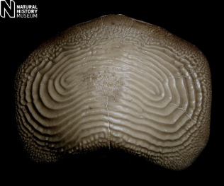
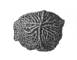
6).
Ptychodus decurrens var. multistriatus - associated teeth - note that the fine ridges link to the sides in crown view to form a concentric pattern (x1.3, Grey
Chalk, Kent, NHMUK (British
Museum (Natural History) London)
PV P2681). Image © 2012 The Natural History Museum, by kind
permission.
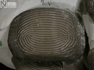
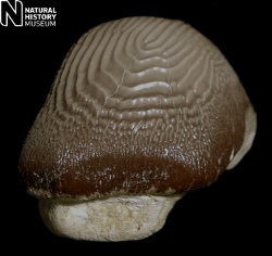
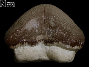
7). Ptychodus decurrens var. multistriatus - (A) Crown (biting surface), (B) lateral (side), and (C) labial views - note that the fine ridges link to the sides in crown view to form a concentric pattern (x1.8, Grey
Chalk, Kent, NHMUK (British
Museum (Natural History) London)
PV P2681). Images © 2012 The Natural History Museum, by kind
permission.
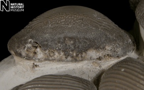
8).
(A,B)
Ptychodus latissimus - (A) oral (biting surface) and (B) side views of a
typical tooth - note gently raised central area with broad pronounced ridges
(x2.1,
Booth Museum, BMB
016995, by kind permission
of John Cooper); (C, D)
Ptychodus latissimus - (A) oral (biting surface) and (B) side views of a
'paucisulcatus'-type tooth - note small number of broad pronounced ridges over central
area, with pronounced triangular profiles (x1.8,
Booth Museum, BMB
016997,
by kind permission of John Cooper).
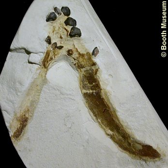
9).
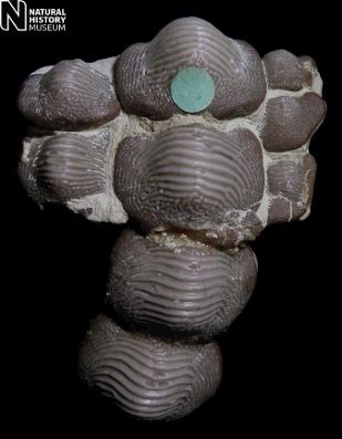
Ptychodus polygyrus - A unique specimen constituting a near complete
association of teeth from the lower jaw, now reconstructed in approximate life
arrangement (Seaford Chalk,
Margate, Kent, Slade & Stilwell Collection, currently on display at
Monkton Nature Reserve);
(A) overview of specimen (x0.7); (B) Detail of teeth from the central row - note
the classic concentric patternation to the ridges of the central area (x1.3);
(C) Detail of tooth in central row showing damage to the tooth (pre-mortem?)
(x4.0).
Images used by kind permission of Ron Stilwell.
10).
Ptychodus polygyrus -
Low angle view of the specimen figure above - note flattened profile to the
teeth (distinguishes Ptychodus polygyrus from Ptychodus concentricus
(see below). Note also that teeth in the central row have well developed
marginal areas, whilst marginal areas are highly reduced to absent in the more
lateral rows (x0.7).
Image used by kind permission of Ron Stilwell.
 A A |
 B B |
11). Ptychodus polygyrus - (A) Crown (biting surface), and (B) lateral views of a very large tooth - note that the relatively coardridges link to the sides to form a concentric patterns(x1.3, Northfleet, Kent, NHMUK (British
Museum (Natural History) London)
PV P33231). Images © 2012 The Natural History Museum, by kind
permission.
 A A |
 B B |
12). Ptychodus polygyrus -
(A) Crown (biting surface), and (B) lateral (side) views of a very
large tooth, associated with the teeth embedded in flint seen below (x1.3, White
Chalk, Grays, Essex, NHMUK (British
Museum (Natural History) London)
PV P12863). Images © 2012 The Natural History Museum, by kind
permission.
13). Ptychodus polygyrus -
associated teeth embedded in flint (x0.8, White
Chalk, Grays, Essex, NHMUK (British
Museum (Natural History) London)
PV P12863). Image © 2012 The Natural History Museum, by kind
permission.
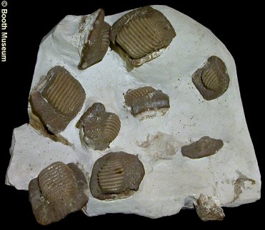
14).
Ptychodus marginalis - a large association of teeth (x0.8, Beachy Head, Sussex, NHMUK (British
Museum (Natural History) London)
PV P6141). Image © 2012 The Natural History Museum, by kind
permission.
 A A |
 B B |
15).
Ptychodus mammillaris; an association of teeth from the Grey Chalk of Sussex
(Willett Collection,
Booth Museum, BMB 007345, by kind permission
of John Cooper); (A) general view of specimen (x0.9); (B) detail (x1.5).
 A A |
 B B |
16).
Ptychodus decurrens; part and counterpart of a unique juvenile specimen,
preserving both the upper and the lower cartilaginous jaws (palatoquadrate
and
Meckel's
cartilage) with accompanying
articulated dentitions. This specimen was the basis for Woodward's much
reproduced Ptychodus reconstruction (1904) (Grey Chalk, Glynde, near
Lewes, Sussex, Willett Collection,
Booth Museum, BMB 008524, by kind permission
of John Cooper); (A) counterpart: oral (biting surface) view of the frontal
portion of the lower dentition and Meckel's cartilage (x1.1); (B) part: ventral / external (underside) view of
the Meckel's cartilage with oral view of the medial portion of upper dentition (x1.1).
 A A |
 B B |
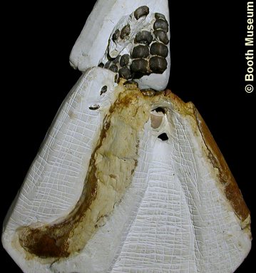
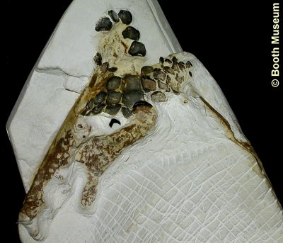
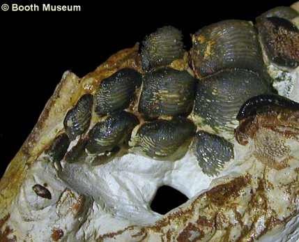
17).
Ptychodus decurrens; more views of the above specimen. (A) dorsal (upperside)
view of the combined part and counterpart. The central and right-hand
portions of cartilage are the palatoquadrate, whilst the left-hand portion is the
Meckel's cartilage. Teeth exposed in oral (biting surface) view (on the left) are
the lower dentition, whilst those exposed in root (underside) view (on the
right) are the upper dentition (x1.1); (B) detail of the lower dentition,
with central row of enlarged teeth on the right. Two teeth of the upper
dentition (root view) are seen far right (x5.0).
 A A |
 B B |
18).
Ptychodus decurrens; more views of the above specimen. (A) oral
(biting surface) view of the medial portion of the upper dentition (x3.5); (B)
oral view of the lower dentition (left hand side of image) and root view of the
upper dentition (right hand side of image) (x2.5).
 A A |
 B B |
19).
Ptychodus decurrens; a unique specimen with the articulated partial lower
and upper dentitions of a moderate sized individual, the front of the animal
presumably lying towards the top of the image. The teeth exposed in oral
(biting surface) view are of the upper dentition, whilst the overlying teeth
exposed in root view are of the lower dentition. The teeth are fused by
pyrite / marcasite (White Chalk of Brighton, Sussex, Willett Collection,
Booth Museum, BMB 008605, by kind permission
of John Cooper); (A) general view of the specimen (x0.8); (B) detail, mainly
showing the left hand side of the upper dentition (x1.5).
 A A |
 B B |
20).
Ptychodus decurrens; more views of the above specimen; (A) Side view,
with the left hand portion of the upper dentition in the fore of the image.
Note that in this image the specimen is in the reverse of life position, with
the biting surface of the upper dentition facing upwards. In this view,
the front of the animal is to the right of the image (x1.0); (B) Detached medial
portion of the lower dentition (x2.0).
 A A |
 B B |
21).
Ptychodus decurrens - A remarkable and seemingly overlooked specimen
preserving the cartilaginous jaws, with the teeth retained in semi-articulation (x1.2,
'Willett Collection',
Booth Museum, BMB 008605 X & XIII, by kind
permission of John Cooper): (A) Overview of the main part of the specimen.
The teeth exposed seem to comprise (mainly? / entirely?) the left and central
portion of the lower dentition. The cartilage bottom-centre of the image
is likely the left-hand
Meckel's
cartilage, whilst the cartilage exposed in section top-right of the image is
questionably the right-hand
palatoquarate
(it is unclear whether the
upper dentition is actually present); (B) The second part of the specimen.
The large tooth seems to have been inserted by the preparator from another
specimen. The cartilage is questionably part of the palatoquadrate.
 A A |
 B B |
22).
Ptychodus decurrens - Details of the above specimen: (A) Detail of the
cartilage (inner surface of left-hand Meckel's cartilage?) showing ornament of
ring-tubercles (x2.5); (B) Detail of the lower(?) dentition (x2.0).
 A A |
 B B |
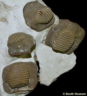
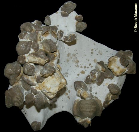
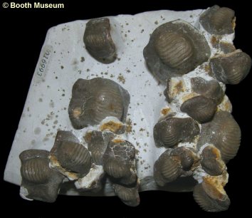
23).
Ptychodus mammillaris - A very large association of teeth (x0.8, Booth Museum, BMB 011272,
016993, by kind permission of John Cooper);
(A) The main part of the association; (B) A further part of the association.
 A A |
 B B |
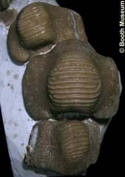
24).
Ptychodus mammillaris - Series of teeth in (A) oral and (B) side views -
note the broad margins and prominently raised central areas crossed by a large
number of relatively fine ridges ((A)
x1.4, (B) x1.6,
Booth Museum, BMB 016999, by kind permission
of John Cooper).
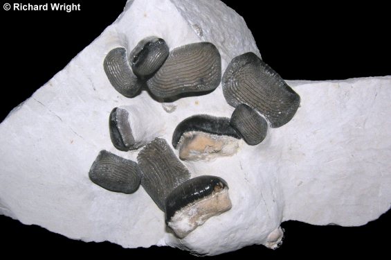
25).
Ptychodus decurrens -
General view of a new specimen (x1.2 Grey Chalk, Bedfordshire, Richard
Wright Collection). Image © 2011 Richard Wright.
 A A |
 B B |
26).
Ptychodus concentricus - (A)
Crown (biting surface) view and (B) lateral (side) views of a large
tooth - note the concentric rings formed by the ridges of the crown,
the crown's sub-conical profile, and highly reduced marginal area /
band (x1.8,
Zig-zag Formation,
Grey Chalk,
Holborough, Kent,
NHMUK (British
Museum (Natural History) London)
PV P10261). Images © 2012 The Natural History Museum, by kind
permission.
 A A |
 B B |
27). Ptychodus concentricus - (A) Lingual
and (B) labial views of a large tooth - note the the crown's sub-conical profile, and highly reduced marginal area / band (x1.8,
Zig-zag Formation,
Grey Chalk,
Holborough, Kent,
NHMUK (British
Museum (Natural History) London)
PV P10261). Images © 2012 The Natural History Museum, by kind
permission.
 A A |
 B B |
28).
Ptychoduc oweni - (A) Crown (biting surface) and (B) lateral (side) views - note that the ridges of the crown radiate
towards the margins, and the diffuse transition to a poorly defined, narrow
marginal area (x2.0,
Zig-zag Formation,
Grey Chalk, Halling,
Kent, NHMUK (British
Museum (Natural History) London)
PV P39125). Images © 2012 The Natural History Museum, by kind
permission.
 A A |
 B B |
29).
Ptychodus mammillaris; oral (biting surface) views of two large teeth; (A)
part of an association from the Grey Chalk of Sussex - see figure 3 below (x2.5, Willett Collection,
Booth Museum, BMB 007345, by kind permission
of John Cooper); (B) isolated tooth sourced from an old collection (x1.7,
Randell Collection, RR1488).
 A A |
 B B |
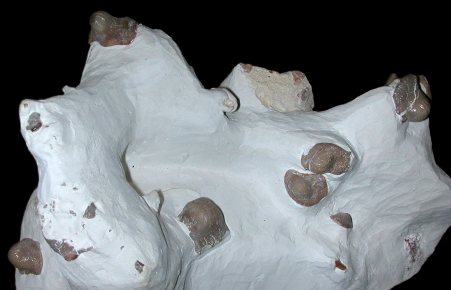
30).
(A) Ptychodus mammillaris; part of an association of teeth (x2.0,
White Chalk?, Burham, Kent, in the collection of
Maidstone Museum, MM 21-622-625, by kind permission of
Ed Jarzembowski); (B) Ptychodus rugosus; detail of an association of
teeth, figured below (x3.0, Belle Tout Marls,
Seaford Chalk,
Seven Sisters, West Sussex, Randell Collection RR1245).
 A A |
 B B |
31).
Ptychodus rugosus; an association of teeth (Belle Tout Marls,
Seaford Chalk,
Seven Sisters, West Sussex, Randell Collection RR1245); (A) general view of
specimen (x0.8); (B) oral surface of a 'nascent' (incompletely developed) tooth
(x3.2).
 A A |
 B B |
32).
Ptychodus rugosus; details of the above association (Belle Tout Marls,
Seaford Chalk,
Seven Sisters, West Sussex, Randell Collection RR1245); (A) x4.0; (B) x3.0.
 A A |
 B B |
33).
Ptychodus cf. mortoni - (A) Crown (biting surface) view and (B)
side
view
profile of an incomplete tooth. This is perhaps the only record of this
form from the UK, but it is well known from the US - Note pointed crown with
ornament of radial ridges passing into a marginal area with granular ornament (x2.9,
Chalk, Winchester area, in the Collections of
Oxford Museum).
Images (A, B) taken from Woodward (1902-1912, Plate LIV,
figures 1 and 1a respectively).
 A
A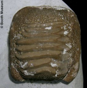 C
C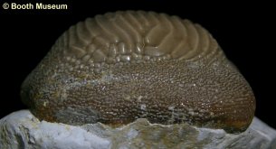 B
B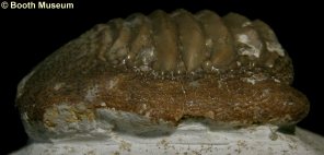 D
D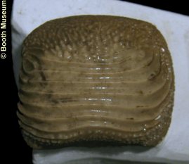
 B
B